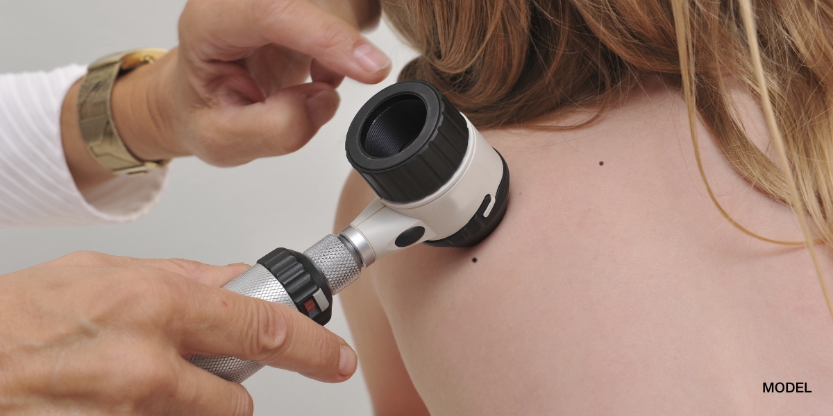
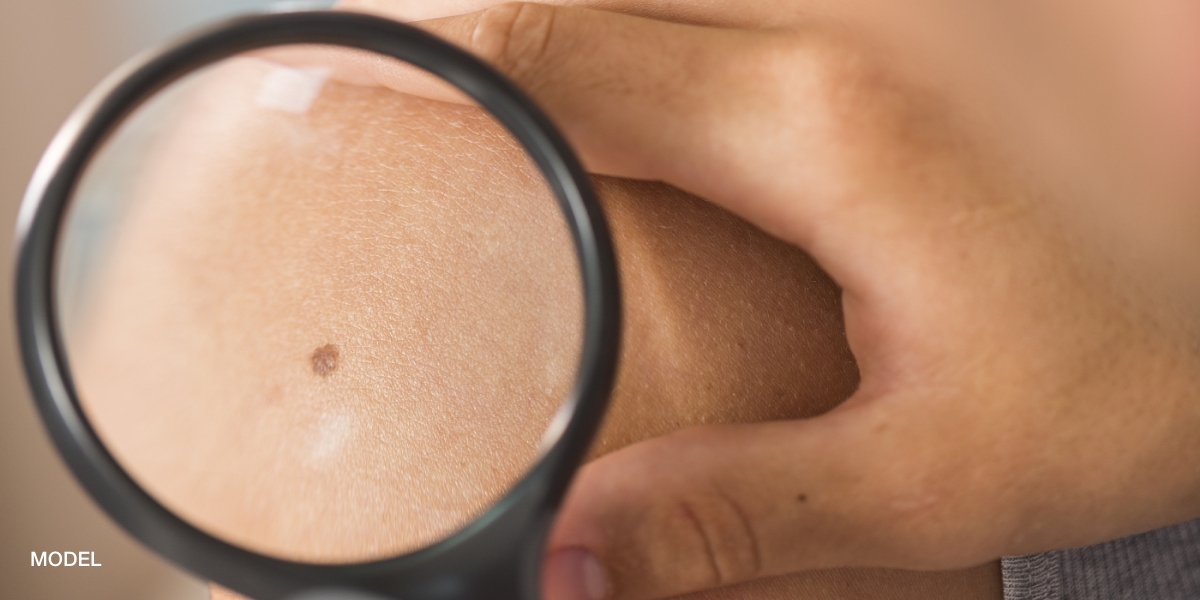
Excisional biopsies are used almost exclusively for skin lesions suspected of being cancer, especially pigmented lesions worrisome for melanoma. As most melanomas are under 6mm, a punch biopsy can be used to obtain an excisional biopsy. Although punch biopsies exist that are 8mm and 1cm, I never use a punch biopsy over 6mm. Punch biopsies create a circular defect and it is impossible to close a circular defect over 6mm as a straight line as the ends of the circle “pooch” forming a raised area called a Burow’s triangle. Thus, an excisional biopsy is best employed for those lesions larger than 6mm. The lesion is excised with a very small margin (1-2mm) so the entire lesion can be viewed by the dermatopathologist. The depth of the biopsy extends to the subcutaneous tissue so it includes all three layers of the skin. I prefer to close the defect with a layer of subcuticular/dermal sutures, which dissolve, followed by a layer of non-absorbable sutures that close the epidermis.
August 3, 2010

Medically reviewed by Anthony J. Perri, M.D.
You May Also Like


Request a Consultation (Sidebar)
Recent Posts
Categories
- Uncategorized (512)
Tags
acne (6)
acne treatment (3)
acne vulgaris (2)
basal cell carcinoma (2)
biopsy (3)
cold urticaria (1)
common skin conditions (11)
dermatologist (15)
dermatology (7)
dr. perri (8)
dry skin (1)
eczema (2)
filiform (1)
health (3)
Herpes (1)
herpessimplex (1)
hives (2)
indentification (1)
keratosis pilaris (1)
Lichen Planopilaris (1)
melanoma (2)
moles (3)
periungual (1)
perri dermatology (10)
prevention (2)
rashes (2)
rosacea (3)
rosacea therapy (2)
skin cancer (6)
skin cancer screening (5)
skin care (2)
skin checks (8)
skin condition (6)
skin conditions (8)
skin damage (2)
skin exam (8)
summertime (3)
sunburn (3)
sunburns (2)
Sunprotection (1)
sunscreen (2)
virus (1)
warts (2)
why perri dermatology (3)
woodlands dermatologist (6)
