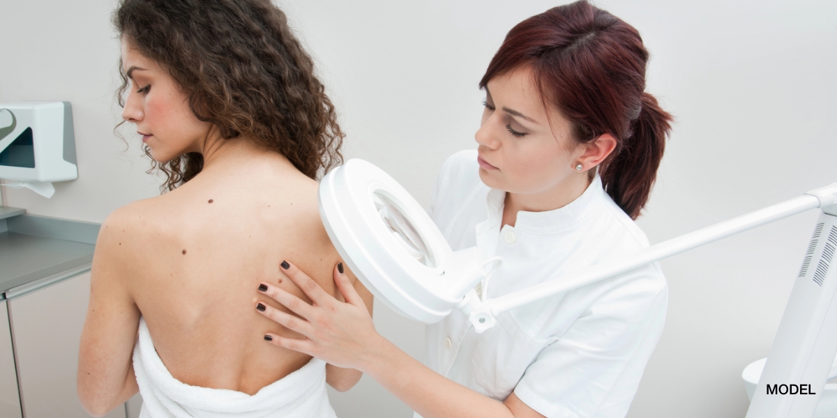
Excision of Basal Cell Cancer is one of the most common methods of treating Basal Cell Cancers and it is the one of the most common methods used in my The Woodlands Dermatology and Conroe Dermatology offices. Excisions of Basal Cell Cancers are usually done in the dermatology clinics in one of our two procedure rooms. Patients are positioned very comfortably on the surgical table and are able to watch one of several relaxing DVDs on a wall mounted flat screen TV. The area to be treated is cleaned with both isopropyl alcohol and Hibiclens to create a sterile field. If the surgical area has hair, this is shaved at the time of surgery prior to creating a sterile field. The area to be treated is diagramed with a sterile marking pen to outline the margins as well as the ellipse. Standard nodular and superficial Basal Cell Cancers require a 5 mm margin of normal appearing skin around what clinically appears to be the full extent of the Basal Cell Cancer. An ellipse is removed rather than a circle since the ends of a circle would not lie flat after suturing. The ellipse elongates the excision along skin tension lines in a 3:1 length to width ratio for optimal surgical cosmesis. A local anesthetic, usually lideocaine 1% with epinephrine is used to anesthetize the surgical area so absolutely no pain or discomfort is felt during the surgery. The patients will feel movement on the surgical area but no pain. Once the ellipse is removed with a surgical scalpel, it is sent to the dermatopathologist for histological inspection of the surgical margins to ensure there are not microscopic “roots” that could not be seen clinically. For approximately 95% of standard Basal Cell Cancers, this 5 mm margin is sufficient to remove the cancer. If the margins are positive, a second excision would be performed several weeks later to remove the remaining cancer cells. Small capillaries are transected during the excision, and these are cauterized with an electrical device called a Hyfrecator. Once hemostasis is achieved, a dermal/subcuticular layer of dissolvable sutures is placed to close the surgical defect. A second “top” layer of non-dissolvable sutures are placed along the incision line which will be removed 1-2 weeks after the surgery. Typically, patients have only mild discomfort after the surgery which is usually alleviated by Tylenol. Vicodin and other narcotic pain medicines are normally not needed. Wound care involves keeping the incision site bandaged with daily wound dressing changes. A large band-aid is normally sufficient to keep the sutures covered. An antibacterial ointment called Bactroban is prescribed at the time of surgery which is to be applied daily to the incision site. Patients are instructed to wash the incision site with an antibacterial soap daily beginning on the second day after surgery and to avoid strenuous activity involving the region of the body with the incision site. Signs of infection are pain/tenderness, swelling, redness and purulent discharge. If a patient notices any of these signs or symptoms they are to call the dermatology clinic immediately. Once the sutures are removed, the scar will remodel over 6 months and its appearance improves in this time period. Typically, the scar will be a thin line that may appear like a natural skin fold or crease. On areas such as the back, scars tend to widen over time due to the thickness and tautness of the skin in this area and this widening is typically unavoidable. Even with clear surgical margins, the surgical area should be examined by a board certified dermatologist every 6-12 months to ensure that no new cancers arise and there is no recurrence of the original cancer as this area is sun damaged enough to produce the original cancer so other cancers may occur in the future.
January 1, 2011

Medically reviewed by Anthony J. Perri, M.D.
You May Also Like


Request a Consultation (Sidebar)
Recent Posts
Categories
- Uncategorized (579)
Tags
acne (6)
acne treatment (3)
acne vulgaris (2)
basal cell carcinoma (2)
biopsy (3)
cold urticaria (1)
common skin conditions (11)
dermatologist (15)
dermatology (7)
dr. perri (8)
dry skin (1)
eczema (2)
filiform (1)
health (3)
Herpes (1)
herpessimplex (1)
hives (2)
indentification (1)
keratosis pilaris (1)
Lichen Planopilaris (1)
melanoma (2)
moles (3)
periungual (1)
perri dermatology (10)
prevention (2)
rashes (2)
rosacea (3)
rosacea therapy (2)
skin cancer (6)
skin cancer screening (5)
skin care (2)
skin checks (8)
skin condition (6)
skin conditions (8)
skin damage (2)
skin exam (8)
summertime (3)
sunburn (3)
sunburns (2)
Sunprotection (1)
sunscreen (2)
virus (1)
warts (2)
why perri dermatology (3)
woodlands dermatologist (6)

