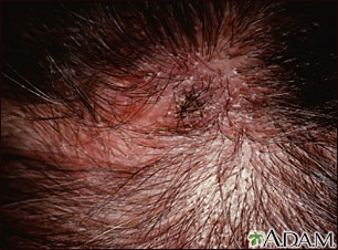
Tinea capitis is a skin condition I encounter in both my Conroe dermatology and The Woodlands dermatology offices. The majority of cases usually occur in children. The most common cause of tinea capitis in the United States is Trichophyton tonsurans. This type of fungus produces a “black dot” type of tinea capitis, in which the hairs are broken off at the level of the scalp causing patches of hair loss with small “black dots” where the hair is severed. The fungus grows either within the hair shaft, endothrix, or outside the hair shaft, ectothrix. Animals can transmit a more inflammatory type of fungus, Microsportum canis, which usually causes hair loss and red scaly plaques on the scalp. These can be associated with lymphadenopathy, lymph node swelling, in which the patient has tender nodules on the base of the neck and behind the ears. In severe cases, a condition called kerion may develop. A kerion is an inflammatory response to the fungus, in which tender red boggy plaques develop in the scalp. These may become co-infected with bacteria and drain pus. The inflammatory response of the kerion can be so severe that the normally transient hair loss of tinea capitis may be permanent as severe scarring can occur. In addition to antifungal and antibacterial medicines, steroids are used to diminish the inflammatory response of a kerion. Tinea capitis can be diagnosed using a KOH sample of the hair and visualizing the fungus under microscopy or through a fungal culture. The oral medicine Lamisi,l is most commonly used to treat tinea capitis, but griseofulvin is another oral option that was used prior to the emergence of Lamisil. Antifungal shampoos such as Nizoral 2% shampoo and Selenium Sulfide 2.5% are also very effective as a topical treatment.
September 20, 2010


