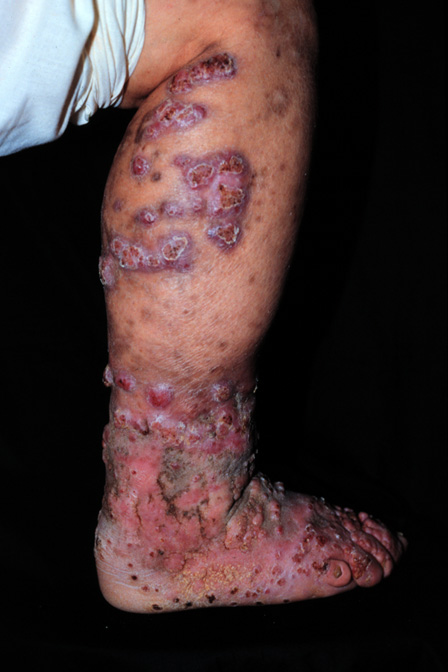
Chromoblastomycosis is a “deep fungus” infection that I occasionally encounter in both my The Woodlands dermatology and Conroe dermatology offices. Chromoblastomycosis is caused by dematiaceous fungi, which are melanin producing, and the most common cause is Fonsecaea pedrosoi. Chromoblastomycosis usually occurs on the lower extremities and is predominantly seen in men. Farmers are the most common group with this fungal infection. The infection usually begins with trauma to the skin such as a thorn piercing the leg. A small wart like papule is the first manifestation of Chromoblastomycosis and gradually increases in size resulting in a large verrucous plaque with scarring. The infection is slowly progressive and only in rare cases does it disseminate to the internal organs. In long standing cases, squamous cell cancers can arise in the Chromoblastomycosis. Diagnosis is made through skin biopsy in which characteristic pigmented fungal sclerotic bodies are seen which appear like”copper pennies.” These “copper pennies” are called Medlar bodies. Surgical treatment or cryotherapy has been effective in smaller lesions, but larger plaques require systemic antifungals such as Itraconazole daily for over a year.
October 24, 2010


