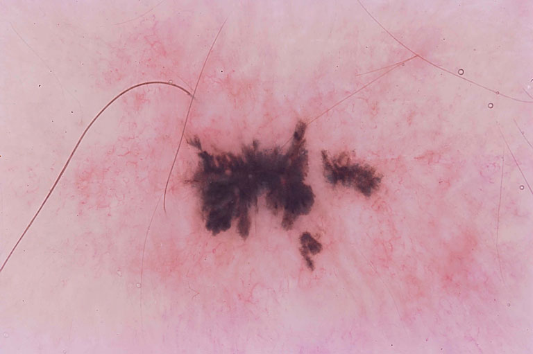
A Recurrent Nevus is a very common skin condition that I encounter on an almost daily basis in both my The Woodlands dermatology and Conroe dermatology offices. A Recurrent Nevus is usually a previously biopsied nevus that ultimately repigments months to years after the original biopsy. In most cases, the biopsy technique was a shave biopsy in which a portion of the nevus was removed for histological evaluation and a remnant of the nevus still remains in the skin. After the normal wound healing process is complete, the remaining melanocytes may produce pigment. The clinical appearance of a Recurrent Nevus is very atypical due to the residual scarring from the previous biopsy and the pigment usually appears very black. Histologically, the scarring causes a pathological appearance with features of melanoma, thus these lesions have been termed “Pseudomelanomas.” Typically, the dermatologist will alert the pathologist that the lesion was previously biopsied so the histological features of scarring with atypical melanocytic proliferation can be investigated to ensure that this melanocytic atypia is confined only to the area above the dermal scar and is not a melanoma but a Recurrent Nevus. Treatment of a Recurrent Nevus depends on the original biopsy results. Nevi arising in scarred areas of the skin can also have the same clinical and histological features of a Recurrent Nevus. In cases of moderate to severe dysplasia, a Recurrent Nevus may be excised with a small surgical margin. 
August 21, 2011


