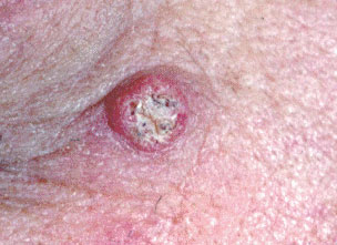
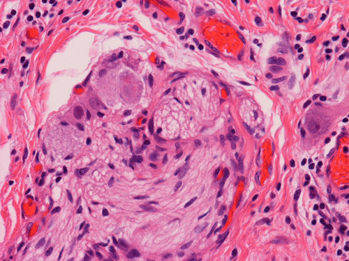
Squamous Cell Cancer can be graded based on the histology of each cancer and is one of the main ways I determine prognosis and management decisions of the Squamous Cell Cancers that I encounter in my The Woodlands and Conroe dermatology offices. Squamous Cell Cancer in situ is the Squamous Cell Cancer version of the Superficial Basal Cell Cancer. Squamous Cell Cancer in situ has not invaded into the epidermis but the cancer cells involve the full thickness of the epidermis. Clinically, Squamous Cell Cancer in situ lesions can become very large and have the appearance of a psoriasis plaque in which they are thick red and scaly. A well differentiated Squamous Cell Cancer, has the best prognosis and is the least aggressive of the Squamous Cell Cancers. Keratoacanthomas are versions of well differentiated Squamous Cell Cancers that arise very quickly and clinically appear like a volcano arising on the surface of the skin. Keratoacanthomas usually arise over 6 weeks, persist for an unspecified time and sometimes spontaneously resolve. On the opposite end of the spectrum, poorly differentiated Squamous Cell Cancers are very aggressive and behave similar to melanomas as they can metastasize rather frequently. Occasionally, perineural invasion is noted on histology of a Squamous Cell Cancer as the cancer cells have infiltrated around nerves in the skin. Perineural invasion also predisposes to metastasis and local recurrence. After a Squamous Cell Cancer that is poorly differentiated or has perineural invasion is excised, adjuvant radiation treatment is usually indicated.


